
This week sees some sunshine and the peak of rich reds, ochres and seasonal yellows here in Piperhill. Strong winds bring down colourful leaf showers. In between the showers, I check all 9 colonies for fondant supplies and replace bags as required. Some bees pay the price and I capture a few in empty fondant bags in the name of education. I freeze them for future dissection: essential for improving microscopy competency.
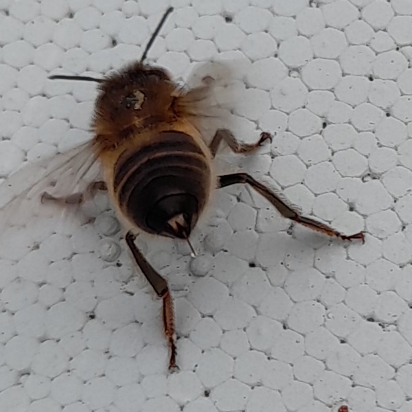
Defensive Behaviour.
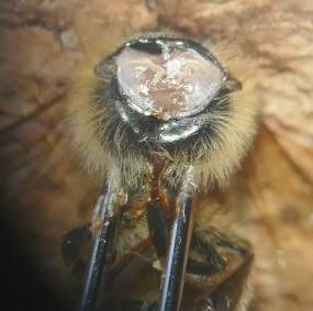
I notice this bee on the roof of a hive I’m about to check for fondant, and it’s not looking well. You can see scarring on its thorax. It’s chilly day to be hanging about though some bees are flying at 4 degrees Celsius. The bee postures like this as I move the roof. If you look closely, you can see that the sting is extended with a drop of venom on the end. I keep well clear of it. Next day I notice the bee dead on the side of the roof.
What About This Behaviour?
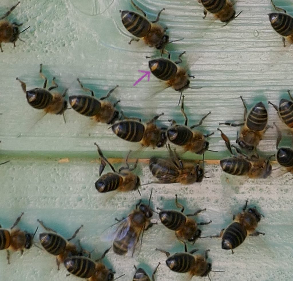
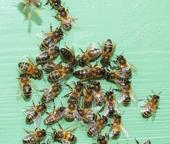
Similar but different behaviour; these bees are busy communicating on the side of the bee shed near the entrance to the observation hive which is full of bees. They are exposing their Nasonov glands, as indicated by the purple arrow, and giving off chemical pheromones to direct their new queen back home after her first mating flight. They are fanning their wings like fury to disperse this chemical message redolent of geraniums.
Searching For Tracheal Mites.
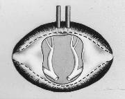
This diagram shows the thoracic collar that must be removed to expose the thoracic tracheae that may be infected with tracheal mites (acarine mites). I remove the heads of 30 dead bees to practice my skills. Removing the collar is not always easy and it may be better to use freshly killed bees. I want to try and get it off in one piece if possible. I will experiment later to compare results, but meanwhile I use the defrosted bees.
I’ve pinned the bee on to a fizzy wine bottle cork, cut at a slant, to hold it stable. The 2 pins secure the thorax to prevent it spinning round as I pull off the collar using the finest tipped forceps I can find. Firstly, to expose the tracheae, I remove the head and first pair of legs by grasping them between the forceps and bending them backwards.
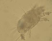
None of the sample bees were infected this week, but a few years ago I found a few live mites crawling about in a piece of tracheae I’d placed on a slide. They are only about 140 microns in length so I needed to magnify the specimen x 400 to see them.
The History.
Scottish scientist Dr John Rennie first described the tracheal mite in 1921 when searching for the cause of the devastating disease that swept the UK wiping out thousands of colonies of honey bees. Radiating out from the Isle of Wight and causing mayhem in commercial apiaries, the condition was named the Isle of Wight Disease. We know now that the tracheal mite was not the cause of this disease and today’s scientists believe that the origin was viral and probably chronic bee paralysis virus.
The resulting disease, devastation and dearth of native British honey bees marked the beginning of importation of honey bees to the UK from other countries, and it prompted Brother Adam (Karl Kerhle) to travel from Buckfast Abbey in search of a better strain of bee. Brother Adam’s world- wide peregrinations led to the conception of the Buckfast bee, but if he had come north to Scotland he would have found native dark bees well suited to our climes and geography. Contrary to the common belief that native dark honey bees were wiped out, Colonsay’s native bee reserve bears testimony to their survival in parts of the country.
Back to Dr Rennie.
Dr Rennie (1865-1928) was Aberdonian and a scientist and graduate from Aberdeen University. His interests were varied and he first worked on finding an early cure for diabetes. He lectured in parasitology at Aberdeen University and was a lead scientist and lecturer at the North of Scotland College of Agriculture at Craibstone. His colleague Bruce White actually discovered the tracheal mite but Rennie identified it naming it Tarsonemus woodi. Later the name would be changed to Acarapis woodi.
The Problems.
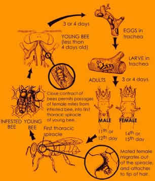
I’ve seen tracheal mites in spring but I don’t think that any of my colonies have been ever been adversely affected by them. The mites enter the first thoracic spiracles of the very young nurse bees where they reproduce in the larger thoracic tracheae. Honey bees like most insects have 10 pairs of spiracles at their sides arranged like the portholes of an ocean liner. These spiracles allow air to enter the respiratory system and they all have valves, apart from the first pair which are the portals of entry for mites.
Honey bees require large intakes of oxygen to power their thoracic flight muscles. They have 3 pairs of thoracic spiracles and 7 abdominal pairs. Oxygen reaches the cells by diffusion through a system of tiny thin walled tubes called tracheoles. Oxygen enters the spiracle and travels through large reinforced tubes called tracheae. They have rings of chitinous material to prevent collapse and these resemble tumble dryer tubing. From the tracheae, oxygen enters large bags called tracheal air sacs which are easily seen on dissection. From there, it travels through the tracheoles throughout the bee body and disperses to all the important muscles, tissues and organs. A mated queen has a spermatheca (sperm storage vessel) covered in a network of fine tracheoles supplying oxygen to keep the sperm alive.
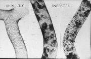
The female mite lays eggs in the thoracic tracheae. The male and female offspring hatch a few days later and develop in a couple of weeks. They mate in the tracheae and the mated female sister comes out of the first spiracle and attaches to a hair. She waits for a young nurse bee to pass and jumps onto her and the cycle continues. Tracheal mites pierce holes in the tracheal walls permanently damaging them as they suck nutritious haemolymph meanwhile clogging up these vital airways and congesting the wing muscles. Affected bees have wings that look dislocated and K-shaped. They cannot fly so crawl up grass stems and die.
Treatment.
There is no recognised treatment here in the UK though varroacides may also kill tracheal mites. Thyme-based Apiguard might be useful but springtime is when you are likely to find mites, after a long winter confinement of bees, but our Scottish temperatures then are not conducive to its efficacy.
Developed Ovaries in Worker Bee.
One last interesting find this week was dissecting a worker bee and finding developed ovaries containing eggs. I tried unsuccessfully to make a slide to show you. However, this is not an uncommon behaviour in worker bees and these eggs are usually policed and removed by other workers who are alerted by chemical signals given off by the eggs. So, I am not unduly worried or tempted to inspect the hive in question. I shall wait till spring and note with interest the situation on first inspection.
Thank you, Ann, I enjoyed reading this beautifully written and illustrated information. x
I’m delighted that you found it interesting knowing your high level of insect knowledge, Margaret Anne. Thank you for commenting.